Wrist Pain & Injuries
What is it?
The wrist is composed of many joints and it is this complex multi-jointed configuration that allows the wrist the kind of movements that we enjoy in our daily activities. Unfortunately, these advantages also predispose the joint to injuries and arthritis. The two most common causes of chronic wrist pain are:
- Scapholunate instability
- Distal radial ulnar joint instability secondary to tear of the triangular fibrocartilage complex (TFCC)
Wrist Sprain
A wrist sprain is an injury to a ligament in the wrist. Ligaments are the connective tissues that connect bones to bones; they could be thought of as the tape that holds the bones together at a joint. These types of injuries are common in falls and sports. The wrist is usually bent backwards when the hand hits the ground. Following the fall, the wrist may be painful with swelling, limited motion and sometimes bruising. Most patients will think nothing of it.
The tell-tale signs that the injuries may be more serious and requires a professional assessment are continued pain for more than a week, especially after a serious fall. Bruising and swelling are serious signs.
SCAPHOLUNATE LIGAMENT INJURY
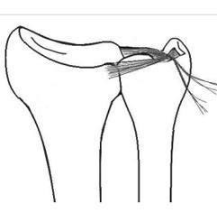
Scapholunate ligament injuries are the most common wrist injuries. This ligament holds the scaphoid and the lunate together. Disruption of this ligament results in scapholunate instability. In the late stages, a gap forms between the scaphoid and lunate bone and is known as scapholunate dissociation.
Symptoms of scapholunate instability include pain, stiffness and swelling. These may be first treated with splinting and non-steroidal anti-inflammatory medicines, and later with cortisone injections. If these treatments fail, surgery may be an option. In the non-arthritic stage, ligament reconstruction is surgery of choice.

MRI showing a tear to the scapholunate ligament and widening of the scapholunate space

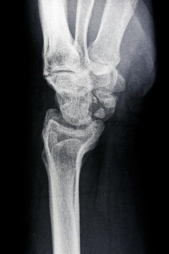
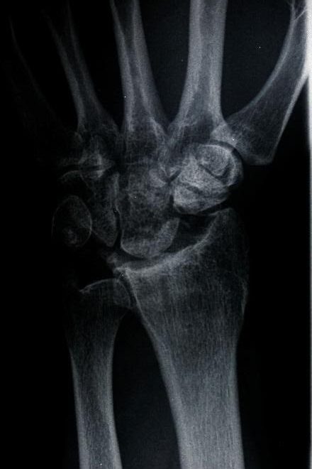
Stressed view of the wrist showing a separation of the scapholunate joint
Stressed view of the wrist showing a separation of the scapholunate joint
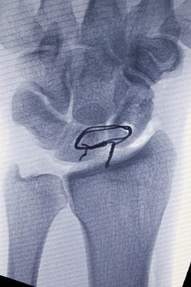
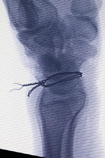
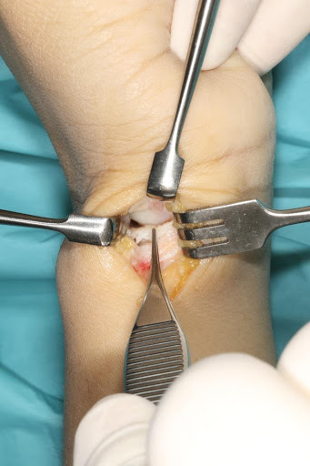
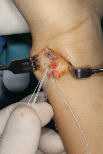
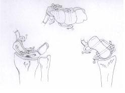
Reconstruct scapholunate of the volar and dorsal ligament with a tendon graft to stabilize the instability is usually the procedure of choice as shown below.
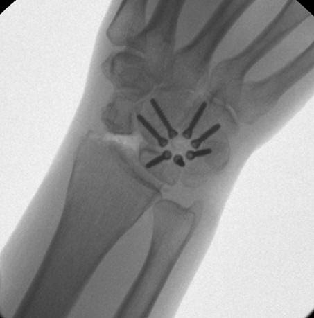
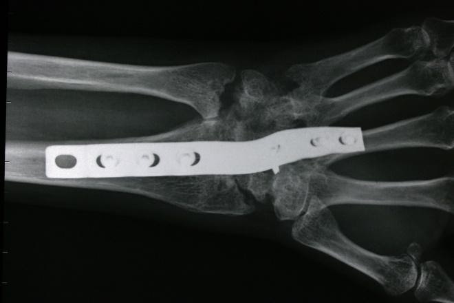
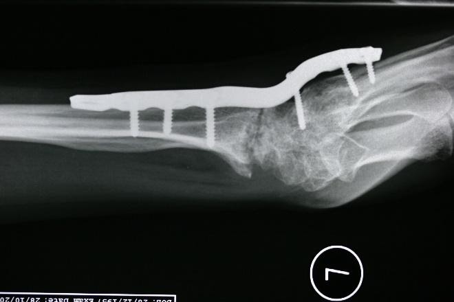
Partial wrist fusion
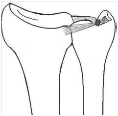
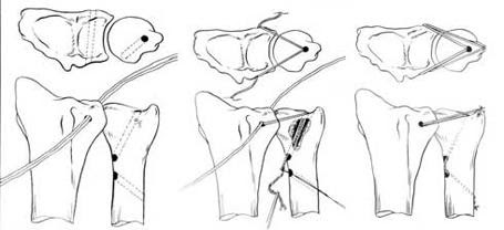
DISTAL RADIAL ULNAR JOINT INSTABILITY AND TEAR OF THE TRIANGULAR FIBROCARTILAGE COMPLEX (TFCC)
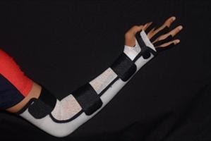
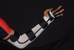
This condition presents with pain over the ulnar aspect of the wrist. Normal wrist usage is usually pain-free. In this condition, the patient experiences pain only when there is a loading on the wrist in a twisting fashion like lifting a heavy luggage bag or when playing tennis. Treatment includes wrist splinting and rest. When the pain settles after splinting, strengthening of the muscles that support the joint may help.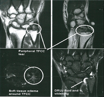
Continued pain after a period of splinting will require further investigations and this may include x-rays and an MRI
 Are you experiencing chronic pain and or continued pain in the wrist? Speak to our hand team at +65 6733 9093. Or book an appointment with our hand specialists for a consultation on your symptoms and treatment options.
Can we help you?

